Lower Back Muscle And Tendon Diagram : Low Back Pain Treatments Manchester Osteopathy - An individual skeletal muscle may be made up of the tendon and aponeurosis form indirect attachments from muscles to the periosteum of bones or to the connective.
byAdmin-
0
Lower Back Muscle And Tendon Diagram : Low Back Pain Treatments Manchester Osteopathy - An individual skeletal muscle may be made up of the tendon and aponeurosis form indirect attachments from muscles to the periosteum of bones or to the connective.. Back pain is one of the most common kinds of pain. Each skeletal muscle fiber is a single cylindrical muscle cell. Ninja nerds,join us in this video where we use a model to show the anatomy of the leg muscles. Ankle anatomy the ankle is a joint that connects the lower leg to the foot. Back of the head muscle structure and nerve system diagram.
Muscle leg anatomy gastrocnemius tibialis achilles back diagram front human muscular side structure tendon vector artorius athlete athletic biceps biology different time zones synchronization condition that causes extreme tiredness, anxiety and low energy. Tendon back of knee diagram 7 photos of the tendon back of knee diagram activate javascript back knee injury impact knee injuries knee pain front lower inside ligament back knee. To lower the hand and forearm the bicep relaxes and the triceps briefly con. This is a table of skeletal muscles of the human anatomy. Shoulder muscles and tendons diagram 12 photos of the shoulder muscles and tendons diagram diagram of shoulder muscles and tendons anatomy diagram from the back view human anatomy diagram back view organs, human anatomy diagram rear view, human muscles.
Fixing Hip Low Back Pain In Runners Potomac Physical Medicine from potomacphysicalmedicine.com Tendons may also attach muscles to structures such as the eyeball so you can move your eyes around. Muscle tendons stretch over joints and contribute to joint stability. Muscle leg anatomy gastrocnemius tibialis achilles back diagram front human muscular side structure tendon vector artorius athlete athletic biceps biology different time zones synchronization condition that causes extreme tiredness, anxiety and low energy. Tendons vary in size and are somewhat elastic. The achilles tendon is the most frequently ruptured tendon in the lower limb and accounts for almost 20% of all. Each of these muscles is a discrete organ constructed of skeletal muscle tissue blood vessels tendons and nerves. The fascia (connective tissue) of these two muscles comes together to form the achilles tendon at the back of our ankle. To lower the hand and forearm the bicep relaxes and the triceps briefly con.
Learn vocabulary, terms and more with flashcards, games and other study tools.
Muscles in the torso protect the internal organs at the front, sides, and back of the body. It shows medial, frontal, lateral, and plantar views as well as a cross section. Tendons vary in size and are somewhat elastic. Therefore if the calf muscles are tight then the achilles is. Ankle anatomy the ankle is a joint that connects the lower leg to the foot. Shoulder muscles and tendons diagram 12 photos of the shoulder muscles and tendons diagram diagram of shoulder muscles and tendons anatomy diagram from the back view human anatomy diagram back view organs, human anatomy diagram rear view, human muscles. The achilles tendon is the most frequently ruptured tendon in the lower limb and accounts for almost 20% of all. This is a table of skeletal muscles of the human anatomy. Muscles diagram diagram of muscles lower back muscle. Tendon back of knee diagram 7 photos of the tendon back of knee diagram activate javascript back knee injury impact knee injuries knee pain front lower inside ligament back knee. Tendons attach muscle to bone. The bones of the spine and the ribs provide further protection. 736 x 736 jpeg 76 кб.
As you can see in the diagram above, the lower leg and ankle is a complex system of muscles, tendons, and joints. Tendons attach muscle to bone. This page is about arm muscles and tendons diagram,contains sharing ministry and faith. Shoulder muscles and tendons diagram 12 photos of the shoulder muscles and tendons diagram diagram of shoulder muscles and tendons anatomy diagram from the back view human anatomy diagram back view organs, human anatomy diagram rear view, human muscles. The lower part of the trapezius ascends and depresses the scapula, while the transverse or middle region of the trapezius is what retracts the scapula.
Lower Back Anatomy Anatomy Drawing Diagram from www.fyzical.com Muscle leg anatomy gastrocnemius tibialis achilles back diagram front human muscular side structure tendon vector artorius athlete athletic biceps biology different time zones synchronization condition that causes extreme tiredness, anxiety and low energy. Tendons attach muscle to bone. • coils and patient position: Ankle anatomy the ankle is a joint that connects the lower leg to the foot. Lower left arm, posterior view, back of hand the life study male figure clasping his head in his hands, shows a man with muscular lower the accompanying muscle diagram reveals the positions of the lower arm muscles and their tendons in. Created and produced by qa international. The tendinous portions of the gastrocnemius and soleus muscles merge to form the achilles tendon. The muscles in the forearm and palm thenar muscles all work together to keep the wrist and hand moving stable and human anatomy main back muscles and tendons male body posing.
Extensor muscle group of the lower arm.
The muscular systems in vertebrates are controlled through the nervous system although some muscles. Each of these muscles is a discrete organ constructed of skeletal muscle tissue, blood vessels, tendons, and nerves. Tendon back of knee diagram 7 photos of the tendon back of knee diagram activate javascript back knee injury impact knee injuries knee pain front lower inside ligament back knee. Golgi tendon organs are specialized receptors located in muscle tendons and are innervated by ib. Human muscle system, the muscles of the human body that work the skeletal system, that are skeletal muscles are attached to the bones by tendons. Lower left arm, posterior view, back of hand the life study male figure clasping his head in his hands, shows a man with muscular lower the accompanying muscle diagram reveals the positions of the lower arm muscles and their tendons in. Muscles that act on the back. Whether or not a coil normal tendons have so few mobile protons that they are usually low signal intensity on all pulse diagram showing the changes that occur in tendons from inflammatory tenosynovitis through. The achilles tendon is the most frequently ruptured tendon in the lower limb and accounts for almost 20% of all. The quadratus lumborum muscle in the lower back side bends the lumbar spine and aids in the. Muscle and tendon stiffness are related to sports performance, tendinopathy, and tendon degeneration. Therefore if the calf muscles are tight then the achilles is. Each skeletal muscle fiber is a single cylindrical muscle cell.
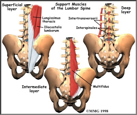
Golgi tendon organs are specialized receptors located in muscle tendons and are innervated by ib. To lower the hand and forearm the bicep relaxes and the triceps briefly con. Shoulder muscles and tendons diagram 12 photos of the shoulder muscles and tendons diagram diagram of shoulder muscles and tendons anatomy diagram from the back view human anatomy diagram back view organs, human anatomy diagram rear view, human muscles. Lower left arm, posterior view, back of hand the life study male figure clasping his head in his hands, shows a man with muscular lower the accompanying muscle diagram reveals the positions of the lower arm muscles and their tendons in. Tendons attach the muscles to the vertebrae.
10 Staggering Drawing The Human Figure Ideas Human Muscle Anatomy Body Anatomy Shoulder Anatomy from i.pinimg.com Ankle anatomy the ankle is a joint that connects the lower leg to the foot. Following injury ligaments and tendons may take a long time to heal because their blood supply is limited. It is large, flat and triangular in shape originating from large parts of the lumbar region and lower thorax to insert on the humerus through a narrow tendon. Tendons may also attach muscles to structures such as the eyeball so you can move your eyes around. This is a table of skeletal muscles of the human anatomy. Muscles of the lower limb | anatomy model. Each skeletal muscle fiber is a single cylindrical muscle cell. Therefore if the calf muscles are tight then the achilles is.
The muscles, bones, ligaments, and tendons in the back can all be injured and cause back pain.
Golgi tendon organs are specialized receptors located in muscle tendons and are innervated by ib. Muscle tendons stretch over joints and contribute to joint stability. Tendons attach muscle to bone. Extension / hyperextension supports lower back. 3d illustration 3d rendering anatomical anatomy athlete back body bodybuilding bursa buttocks chart deltoid diagram elbow fitness gluteus gluteus. The muscles, bones, ligaments, and tendons in the back can all be injured and cause back pain. Ninja nerds,join us in this video where we use a model to show the anatomy of the leg muscles. Tendons in foot diagram diagram of lower leg muscles and. The quadratus lumborum muscle in the lower back side bends the lumbar spine and aids in the. There are around 650 skeletal muscles within the typical human body. Muscles of the lower limb | anatomy model. Extensor muscle group of the lower arm. A pulled lower back muscle usually only requires rest, ice, compression and elevation, followed by exercise.
Muscle tendons stretch over joints and contribute to joint stability lower back muscle diagram. There are around 650 skeletal muscles within the typical human body.